Genetic and Hereditary Risk in Age-Related Macular Degeneration (AMD)
This study guide provides a comprehensive review of the molecular mechanisms, genetic predispositions, and environmental interactions associated with Age-Related Macular Degeneration (AMD). It is designed to facilitate a deep understanding of the hereditary risks, protective alleles, and emerging personalized therapeutic strategies in the field of ophthalmic genomics.
Part 1: Short-Answer Quiz
Instructions: Answer the following ten questions in 2-3 sentences based on the provided research.
- What are the primary clinical and pathological differences between the "dry" and "wet" phenotypes of advanced AMD?
- How does the Y402H polymorphism in the CFH gene increase the risk of developing AMD?
- What specific cellular and mitochondrial abnormalities are observed in RPE cells harboring high-risk CFH genotypes?
- Explain the genetic-environmental synergy observed between smoking and the CFH CC genotype.
- What is the core of the scientific controversy regarding the chromosome 10q26 locus?
- How does the overexpression of the HTRA1 protease physically damage the eye's anatomy?
- Describe the role of the ARMS2 gene in maintaining mitochondrial health via mitophagy.
- How do Polygenic Risk Scores (PRS) improve the predictive accuracy of clinical models for AMD?
- Contrast the impact of the APOE ε4 and ε2 alleles on AMD risk and clinical onset.
- According to metabolomic research, how does the systemic "signature" of the blood change as AMD progresses to its late stage?
Part 2: Quiz Answer Key
- Dry AMD is characterized by geographic atrophy (GA), which involves the wasting away of macular photoreceptors and the retinal pigment epithelium (RPE). In contrast, wet AMD involves choroidal neovascularization (CNV), where abnormal blood vessels break through Bruch’s membrane, leading to hemorrhage, exudation, and rapid scarring.
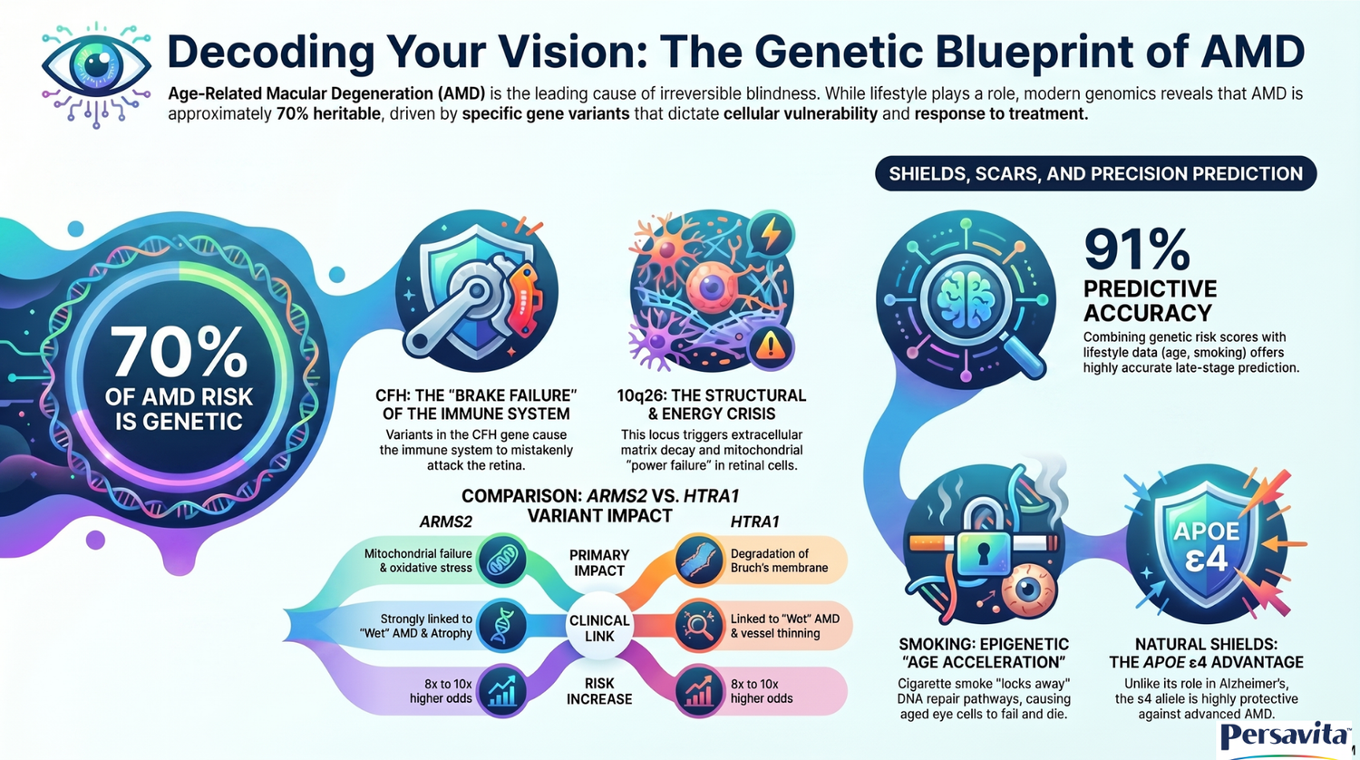
- The Y402H variant involves a tyrosine-to-histidine substitution that reduces the affinity of the CFH protein for protective ligands on Bruch’s membrane and the RPE. Without this anchoring, CFH cannot inhibit the alternative complement pathway, leading to unchecked inflammation, C3 convertase formation, and tissue damage via the membrane attack complex (MAC).
- Cells with the high-risk CFH genotype exhibit decreased mitochondrial mass and density, reduced electron transport chain proteins, and lower ATP synthesis. Additionally, these cells show elevated mitochondrial DNA damage and a diminished capacity to handle oxidative stress.
- Smoking significantly exacerbates the bioenergetic deficits of the high-risk CFH genotype, causing a dramatic drop in cell viability and accelerated mitochondrial decay. Epidemiologically, this results in a 34-fold increased odds ratio for late-stage AMD in smokers who are homozygous for the CFH CC genotype compared to non-smokers with the low-risk genotype.
- The 10q26 locus contains three closely linked genes (PLEKHA1, ARMS2, and HTRA1) that are inherited together due to high linkage disequilibrium. Researchers debate whether HTRA1 (via extracellular matrix degradation) or ARMS2 (via mitochondrial and mitophagy dysfunction) is the primary causal driver of the disease.
- Excess HTRA1 cleaves critical structural proteins like fibulin 5 and ApoE, leading to the fragmentation of the elastic layer of Bruch's membrane. This degradation causes the membrane to thicken and collagen fibrils to loosen, which facilitates the invasion of abnormal choroidal blood vessels (CNV).
- The ARMS2 protein is concentrated in the mitochondria, where it helps regulate mitophagy, the process of clearing damaged mitochondria. The high-risk A69S variant impairs this process and reduces superoxide dismutase (SOD) activity, leading to the accumulation of reactive oxygen species (ROS) and dysfunctional RPE cells.
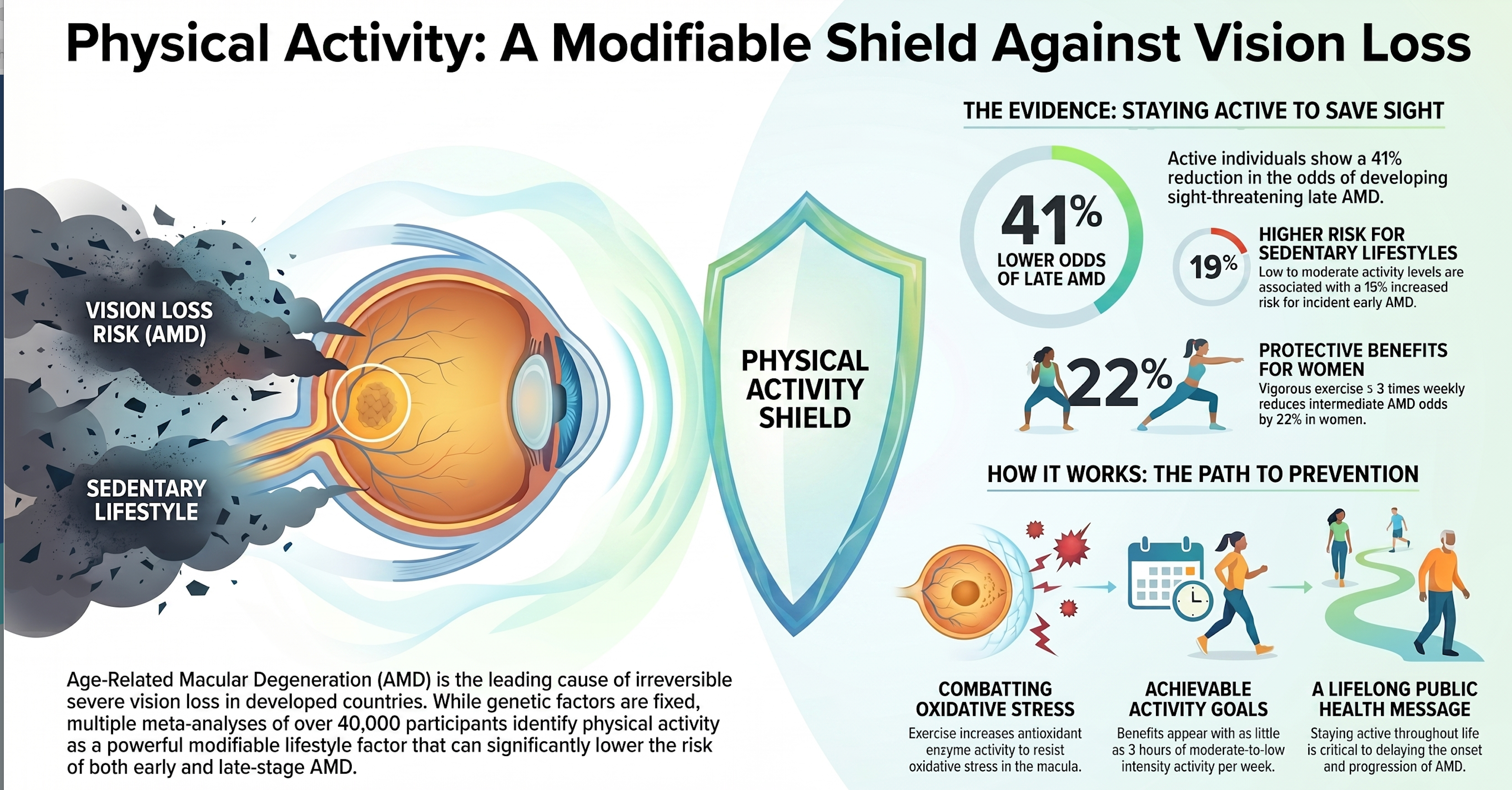
- While traditional models using only non-genetic factors (age, smoking, etc.) have an predictive accuracy (AUC) of 79%, integrating updated PRS models increases this to 91%. This allows for the identification of high-risk individuals before vision loss occurs and helps predict which patients will respond best to complement-targeted therapies.
- The ε4 allele is highly protective, halving the risk of advanced AMD because the ApoE4 protein has high molecular mobility that helps clear lipid waste from Bruch's membrane. The ε2 allele increases risk and can accelerate the clinical diagnosis of wet AMD by up to 4.7 years.
- Early stages are dominated by lipid alterations, such as increased large HDL subparticles and decreased VLDL. Late-stage AMD shifts toward amino acid depletion (e.g., tyrosine, histidine) and an elevation of ketone bodies, suggesting systemic energetic strain and fatty acid oxidation.
Part 3: Essay Questions
Instructions: Use the provided source context to develop detailed outlines or responses for the following prompts. (Answers not provided).
- The Complement-Metabolic Intersection: Analyze how a single genetic variant, such as CFH Y402H, can simultaneously disrupt immune regulation and cellular bioenergetics. Discuss the implications this has for developing multi-target therapeutics.
- Environmental Epigenetics: Using the Johns Hopkins research on cigarette smoke, explain how environmental stressors "lock away" DNA repair pathways in aged RPE cells and why young cells are better equipped to survive these stressors.
- Precision Medicine in Ophthalmology: Evaluate the current evidence for personalizing AMD treatments based on genetic stratification. Focus on complement inhibitors for high-PRS patients and protease inhibitors for those with 10q26 variants.
- The Role of Lipids in Retinal Health: Compare the biological mechanisms of ABCA1, APOE, and LIPC in maintaining Bruch's membrane. Explain how failures in these lipid-processing pathways lead to the formation of drusen.
- Innate Protection: Examine the mechanisms of protective alleles like PELI3 A307V and CFHR1/CFHR3deletions. Discuss how these variants offer "innate retinal shields" by modulating the immune response.
Part 4: Comprehensive Glossary
|
Term |
Definition |
|
Alternative Complement Pathway |
A self-amplifying arm of the innate immune system that clears debris and pathogens; its dysregulation is a primary driver of AMD. |
|
ARMS2 |
Age-Related Maculopathy Susceptibility 2; a protein localized to mitochondria and the ECM involved in mitophagy and debris clearance. |
|
Bruch’s Membrane (BrM) |
The thin, multi-layered extracellular matrix between the RPE and the choroid that acts as a metabolic sieve for the retina. |
|
C3 Convertase |
An enzyme complex (C3bBb) formed during complement activation that cleaves C3 into inflammatory mediators. |
|
Choroidal Neovascularization (CNV) |
The growth of abnormal, leaky blood vessels from the choroid into the retina; the hallmark of "wet" AMD. |
|
Complement Factor H (CFH) |
The primary soluble inhibitor of the alternative complement pathway; it protects host tissues from bystander immune damage. |
|
Drusen |
Extracellular deposits of protein and lipid that accumulate beneath the RPE; a clinical sign of early-to-intermediate AMD. |
|
Epigenetics |
Modifications to gene expression (like DNA methylation or chromatin changes) that do not alter the underlying DNA sequence. |
|
Geographic Atrophy (GA) |
The advanced form of "dry" AMD characterized by the progressive loss of RPE cells and photoreceptors. |
|
HTRA1 |
High-Temperature Requirement A Serine Peptidase 1; an enzyme involved in ECM remodeling whose overexpression degrades Bruch's membrane. |
|
Linkage Disequilibrium (LD) |
The non-random association of alleles at different loci, meaning they are inherited together more often than expected by chance. |
|
Macula |
The central region of the retina responsible for high-acuity color vision and sharp central perception. |
|
Membrane Attack Complex (MAC) |
The final product of the complement cascade (C5b-9) that forms pores in cell membranes, leading to cell lysis. |
|
Mitophagy |
The selective autophagic clearance of damaged or dysfunctional mitochondria to maintain cellular health. |
|
Polygenic Risk Score (PRS) |
A single numerical value that aggregates the cumulative effect of many minor and major genetic variants to predict disease risk. |
|
Retinal Pigment Epithelium (RPE) |
A layer of cells behind the photoreceptors essential for visual cycle maintenance, waste clearance, and nutrient transport. |
|
Single-Nucleotide Polymorphism (SNP) |
A variation at a single position in a DNA sequence among individuals. |
|
Superoxide Dismutase (SOD) |
An enzyme that repairs cells and reduces damage caused by superoxide, the most common free radical in the body. |
Leave a comment
This site is protected by hCaptcha and the hCaptcha Privacy Policy and Terms of Service apply.